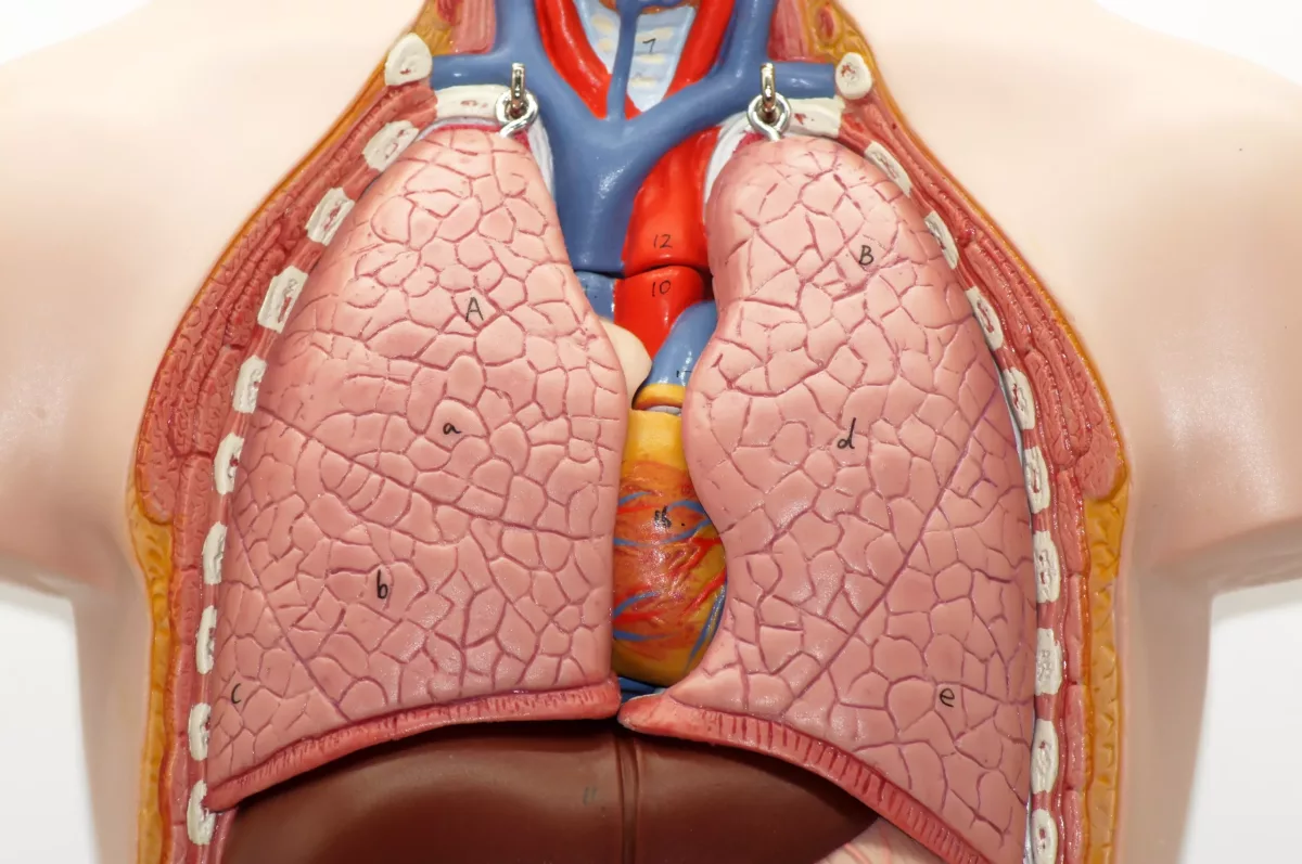
A narrowing of the main artery in the body (aorta) is called aortic coarctation. Therefore, this condition makes the heart work harder to supply the body’s needs with blood. In most cases, this condition is present at birth (congenital heart defect), but in some people, it may occur later in life. Commonly, this condition happens along with other congenital heart defects.
In most cases, the condition is successfully treated. Anyway, regular heart checkups are recommended to watch for heart health changes.
Symptoms
The symptoms of this disease depend on how narrowed the artery is. Most people with this condition do not have symptoms, and their hearts seem to be healthy. In more severe cases, when the aorta is extremely narrowed, the symptoms may appear right after birth. Check below some symptoms of coarctation of the aorta in infants:
- Irritability
- Skin color changes
- Heavy sweating
- Feeding difficulties
- Breathing problems
However, it may cause symptoms later in life as well. These include:
- Nosebleeds
- Cold feet
- Leg cramps
- Hypertension (high blood pressure)
- Chest pain
- Muscle weakness
- Headaches
It is advised to go to the nearest emergency room or call 911 in the U.S. if any of the following symptoms appear. Examples include:
- Fainting
- Sudden shortness of breath
- High blood pressure that occurs without any obvious reason
Causes
Healthcare providers do not fully understand why coarctation of the aorta occurs, but it is commonly present at birth. Generally, a congenital heart defect happens during pregnancy in the womb. The exact cause of most congenital heart defects cannot be identified.
In rare cases, this congenital heart defect may occur later in life. Check below some factors that may trigger the condition:
- Traumatic injuries
- Extreme cholesterol and fat buildup in the arteries (also known as atherosclerosis)
- Swelling and irritation of the heart’s blood vessels (also known as Takayasu arteritis)
Risk Factors
While the exact cause of this congenital heart defect cannot be determined, physicians identified some factors that could increase the risk of developing it. For example:
- Male sex
- Genetic disorders (such as Turner syndrome)
- Other congenital heart defects (heart disease present at birth)
Check below some examples of congenital heart defects often associated with aorta coarctation:
- Bicuspid aortic valve – This valve is located between the aorta (the main body’s artery) and the left lower heart chamber. A bicuspid aortic valve occurs when this valve has two flaps (also called cusps) instead of three.
- Subaortic stenosis – This condition happens when the area below the aortic valve is narrowed. Therefore, it blocks the flow of blood from the lower left heart chamber to the main artery of the body.
- Patent ductus arteriosus – A blood vessel that connects the left lung artery to the aorta is called the ductus arteriosus. In normal circumstances, the ductus arteriosus closes after birth, but in people with this condition, it remains open.
- Holes in the heart – There are people with aorta coarctation who are born with a hole in the heart. When a hole happens in the upper heart’s part, the condition is called an atrial septal defect, but when in the lower part of the heart, it is called a ventricular septal defect.
- Congenital mitral valve stenosis – This is a valve disease type that is present at birth. It causes the valve between the upper and lower left heart chambers to narrow, which causes the blood to move more forcefully through the valve.
What Are The Potential Complications of Coarctation of the Aorta?
Complications of this congenital heart defect usually occur because the lower left heart chamber works harder to pump blood through a narrowed artery. As a result, blood pressure goes to the lower left chamber of the heart. Furthermore, aorta coarctation may cause the heart chamber wall to become thick (ventricular hypertrophy). Check below some complications:
- Chronic (long-term) high blood pressure (also called hypertension)
- Brain aneurysm (a weakened or bulging artery in the brain)
- Bleeding in the brain
- A tear or rupture in the aorta (also known as aortic dissection)
- Aortic aneurysm (a bulge in the aorta wall)
- Coronary artery disease
- Stroke
Without treatment, it may lead to life-threatening complications, including heart or kidney failure and even death.
Discuss with your healthcare professional about ways to prevent aortic coarctation complications.
How to Prevent Coarctation of the Aorta?
Unfortunately, there are no sure ways to prevent this congenital heart defect. However, you can take steps to reduce the risk. Consult with your doctor for more details.
Diagnosis
Diagnosis of this congenital heart defect depends on how severe it is. For instance, in severe cases, it is diagnosed right after birth. Sometimes, it can be identified during pregnancy with an ultrasound. In mild cases, the disease may not be found until it causes symptoms.
Commonly, during diagnosis, physicians measure the blood pressure in arms and legs, evaluate your medical history, and ask some questions about the symptoms. They also perform some tests that help confirm the disease and rule out others that cause similar symptoms.
Tests
- Echocardiogram – This test uses sound waves to make images of the beating heart and how blood flows through the heart muscle. It may show how narrow the aorta is.
- Electrocardiogram (EKG or ECG) – It is a quick and painless test that measures the heart’s electrical activity. If the aorta is severely narrowed, the ECG may show thickening of the lower chambers of the heart walls.
- Chest X-ray – This is an imaging test that makes images of the heart and lungs. It may show coarctation of the aorta.
- MRI (magnetic resonance imaging) and CT (computerized tomography) scans – These are other imaging tests that make detailed pictures of different body structures and organs. They can also identify the exact location of the aorta coarctation.
- Coronary angiogram with cardiac catheterization – During this test, doctors use a long and flexible tube that is usually inserted through a major blood vessel (often in the groin or wrist) to deliver dye. Thereafter, doctors perform an X-ray test to check for abnormalities linked with the disease.
- CT angiogram – It also involves a dye and special X-rays to check the heart’s blood vessels (also known as coronary arteries).

Treatment
The treatments for people with coarctation of the aorta are usually different because they depend on the severity of the disease, existing health problems, age, preferences, and other factors. However, physicians commonly recommend medications, heart therapies, or surgery.
Medicines
- Antihypertensives – This group of medications is prescribed by doctors to control blood pressure before repair surgery. While surgery can improve blood pressure, most people require blood pressure medicines after repair.
- Medicines to maintain the ductus arteriosus open – In normal circumstances, the ductus arteriosus closes after birth. Physicians may prescribe some medications to keep it open until the coarctation of the aorta is repaired.
Other Treatments
The following procedures are usually used by doctors to repair the aorta coarctation. Examples include:
- Balloon angioplasty and stenting – Commonly, this is the primary treatment for people with aortic coarctation. It helps widen the aorta and improve blood flow. During this procedure, surgeons will use a small tube (catheter) and a small balloon to open the narrowed blood vessel.
- Resection with end-to-end anastomosis – During this surgery, they will remove the narrowed part of the aorta. After that, surgeons connect two healthy aorta parts (also known as anastomosis).
- Subclavian flap aortoplasty – This procedure involves taking a part of the blood vessel that delivers blood to the left arm (left subclavian artery) and using it to widen the aorta.
- Bypass graft repair – This surgery is often performed to make an additional pathway for blood to flow around the narrowed aorta.
- Patch aortoplasty – It is a surgery that uses pieces of material to widen the artery. This procedure is effective if the coarctation affects the long part of the aorta.
Frequently Asked Questions
What happens in coarctation of the aorta?
In people with this condition, a part of the body’s main artery (aorta) becomes narrowed. As a result, the heart muscle works harder to pump blood for the body’s needs. In most cases, this condition is present at birth (also called a congenital heart defect).
What is the life expectancy of a person with aortic coarctation?
Generally, the life expectancy is approximately 35 years. As per studies, most people with this congenital heart defect die before 50 years old.
What are the long-term effects of coarctation of the aorta?
People with this condition may experience some complications, even with successful treatment. Check below some examples:
- Aneurysm
- Increased risk of coronary and cerebral artery disease
- Recoarctation
- Hypertension (high blood pressure)
- Exercise intolerance
- Aortic valve disease
- Left ventricular dysfunction
This article does not contain a complete list of aorta coarctation complications. Ask your healthcare professional if you have additional questions.


